摘要详情
56 / 2021-10-14 21:53:20
A preliminary study of real-time imaging for MR guided vascular intervention and image quality assessment
Interventional MRI,real-time imaging sequences,3D printed,image quality
终稿
Background: Interventional MRI have significant benefits for image-guided intravascular intervention and related vascular procedures. Real-time MRI could provide a radiation-free alternative to X-ray guidance of cardiovascular catheterization, enables superb tissue contrast depiction in multi-contrast imaging without administration of contrast agent. In this study, we evaluated the image quality of two MR real-time sequences, fast low angle shot (FLASH) and true fast imaging with steady-state precession (True FISP), for MR guided vascular intervention using MR compatible guidewire and balloon in a 3D printed aorta phantom.
Methods: In this study, an MR guided intervention system were setup, which consisted of a 3T scanner (MAGNETOM Skyra, Siemens Healthcare, Erlangen, Germany) and MRI compatible monitor (Fuqing Medical Technology Co., Ltd, Hefei, Anhui, China). All experiments were performed using a peripheral balloon catheter (PTA35-6040B, DK MEDTECH, Suzhou, Jiangsu, China), which filled with 0.8ml 1% Gadodiamide mixed with normal saline and magnetic resonance compatible guidewire (diameter=0.035 in, length=150cm) (EPflex, Dettingen an der Erms, Germany). Guidewire consisted of a high-strength para-aramid synthetic fiber core, surrounded by a bending-resistant high-performance polymer and a hydrophilic coating, the markers in the guidewire were discretely mosaicked for navigation. All experiments were performed in a 3D printed aorta phantom printed by 3D printer (SPS250j, Shaanxi Hengtong Intelligent Machine Co., Ltd, Shaanxi, China.). The aorta phantom was made of transparent resin with CTA data from a normal adult (Fig 1). The interventional procedure was guided and recorded by FLASH and True FISP. The parameters of the real-time imaging sequence were: (1) FLASH, TR/TE =6.8/2.65ms, FA=12º, FOV=360×147mm, Slice thickness=10mm, Phase partial Fourier=6/8, bandwidth=230Hz/Px, (2) True FISP, TR/TE =321.35/1.88ms, FA=49º FOV=360×144mm, Slice thickness=10mm, bandwidth=1116Hz/Px.
The real-time images were evaluated objectively and subjectively. For objective assessment, the image signal-to-noise ratio (SNR), contrast noise ratio (CNR), geometric distortion, and image uniformity of two sequences were measured. The signal intensity and noise at different positions of the image were measured and their mean value was taken to calculate the SNR. The SNR was calculated by:
SNR=Signalnormal saline/SDnoise.
The CNR between gadolinium-filled balloon and normal saline in the phantom was measured. The CNR was calculated by:CNR=|Signalballoon-Signalnormal saline|/SDnoise.
Regarding image uniformity, most areas of multiple images were measured, and the mean value of their uniformity was calculated. The image uniformity was calculated by:U={1-(Signalmax-Signalmin/Signalmax+Signalmin)}*100%.
In terms of geometric distortion, the actual length of the gadolinium balloon and its length in the image was measured, and then the geometric distortion was calculated by:GD=|Lengthimage-Lengthactual|/Lengthimage*100%.
For subjective assessment, Likert scoring was applied to assess the image sharpness, image distortion, and artifacts of all sequences. The images quality was scored subjectively by two experienced radiologists with more than six-year practice. The evaluation criteria were as follows: 1 points = poor image sharpness with serious distortion or artifacts, 2 points = medium image sharpness with medium distortion or artifact, 3 points = good image sharpness with slight distortion or artifacts, 4 points = excellent image sharpness with no distortion or artifacts.
The results of the objectively and subjectively analyses were compared between the two sequences using independent sample T-test by IBM SPSS Statistics 26.
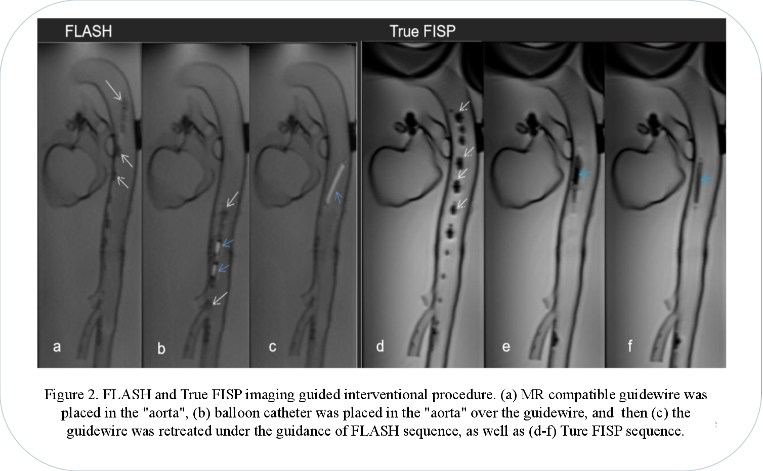
Results: Both sequences temporal resolution was 2 frames/s and spatial resolution was 1.1×1.1×10mm (Fig 2). For objective assessment the two sequences (FLASH vs True FISP) showed significant different SNR (102±16 vs 618±137, p<0.05) and CNR (73.5±18 vs 162.2±46, p<0.05). Image uniformity (80%±6% vs 74%±4%, p=0.133) and geometric distortion (2%±1%, 2%±1%, p=0.717) did not showed significant difference. Two radiologists reached agreement on subjective scoring. The subjective scores of two sequences did showed significant difference, and the FLASH score was higher than the True FISP score (FALSH=3.7±0.5, True FISP=3.1±0.9, p<0.05).
The artifacts of the guidewire were moderate for FLASH, and the balloon was high signal. In True FISP, the artifacts of the guidewire were severe, the balloon was low signal and blurred by the artifacts of the guidewire.
Discussion: Both sequences have sufficient SNR, CNR, image uniformity and the degree of geometric distortion was also within the acceptable range. The FLASH sequence has sufficient guiding ability in the actual application process, the artifacts of the guidewire were moderate. Although the SNR and CNR were higher for True FISP, the artifacts of the guidewire were severe, and blurred display of balloon.
Conclusions: In conclusion, the study demonstrates the MR real-time imaging, FLAH sequence, is feasible of guiding cardiovascular interventions.
Methods: In this study, an MR guided intervention system were setup, which consisted of a 3T scanner (MAGNETOM Skyra, Siemens Healthcare, Erlangen, Germany) and MRI compatible monitor (Fuqing Medical Technology Co., Ltd, Hefei, Anhui, China). All experiments were performed using a peripheral balloon catheter (PTA35-6040B, DK MEDTECH, Suzhou, Jiangsu, China), which filled with 0.8ml 1% Gadodiamide mixed with normal saline and magnetic resonance compatible guidewire (diameter=0.035 in, length=150cm) (EPflex, Dettingen an der Erms, Germany). Guidewire consisted of a high-strength para-aramid synthetic fiber core, surrounded by a bending-resistant high-performance polymer and a hydrophilic coating, the markers in the guidewire were discretely mosaicked for navigation. All experiments were performed in a 3D printed aorta phantom printed by 3D printer (SPS250j, Shaanxi Hengtong Intelligent Machine Co., Ltd, Shaanxi, China.). The aorta phantom was made of transparent resin with CTA data from a normal adult (Fig 1). The interventional procedure was guided and recorded by FLASH and True FISP. The parameters of the real-time imaging sequence were: (1) FLASH, TR/TE =6.8/2.65ms, FA=12º, FOV=360×147mm, Slice thickness=10mm, Phase partial Fourier=6/8, bandwidth=230Hz/Px, (2) True FISP, TR/TE =321.35/1.88ms, FA=49º FOV=360×144mm, Slice thickness=10mm, bandwidth=1116Hz/Px.
The real-time images were evaluated objectively and subjectively. For objective assessment, the image signal-to-noise ratio (SNR), contrast noise ratio (CNR), geometric distortion, and image uniformity of two sequences were measured. The signal intensity and noise at different positions of the image were measured and their mean value was taken to calculate the SNR. The SNR was calculated by:
SNR=Signalnormal saline/SDnoise.
The CNR between gadolinium-filled balloon and normal saline in the phantom was measured. The CNR was calculated by:CNR=|Signalballoon-Signalnormal saline|/SDnoise.
Regarding image uniformity, most areas of multiple images were measured, and the mean value of their uniformity was calculated. The image uniformity was calculated by:U={1-(Signalmax-Signalmin/Signalmax+Signalmin)}*100%.
In terms of geometric distortion, the actual length of the gadolinium balloon and its length in the image was measured, and then the geometric distortion was calculated by:GD=|Lengthimage-Lengthactual|/Lengthimage*100%.
For subjective assessment, Likert scoring was applied to assess the image sharpness, image distortion, and artifacts of all sequences. The images quality was scored subjectively by two experienced radiologists with more than six-year practice. The evaluation criteria were as follows: 1 points = poor image sharpness with serious distortion or artifacts, 2 points = medium image sharpness with medium distortion or artifact, 3 points = good image sharpness with slight distortion or artifacts, 4 points = excellent image sharpness with no distortion or artifacts.
The results of the objectively and subjectively analyses were compared between the two sequences using independent sample T-test by IBM SPSS Statistics 26.
Results: Both sequences temporal resolution was 2 frames/s and spatial resolution was 1.1×1.1×10mm (Fig 2). For objective assessment the two sequences (FLASH vs True FISP) showed significant different SNR (102±16 vs 618±137, p<0.05) and CNR (73.5±18 vs 162.2±46, p<0.05). Image uniformity (80%±6% vs 74%±4%, p=0.133) and geometric distortion (2%±1%, 2%±1%, p=0.717) did not showed significant difference. Two radiologists reached agreement on subjective scoring. The subjective scores of two sequences did showed significant difference, and the FLASH score was higher than the True FISP score (FALSH=3.7±0.5, True FISP=3.1±0.9, p<0.05).
The artifacts of the guidewire were moderate for FLASH, and the balloon was high signal. In True FISP, the artifacts of the guidewire were severe, the balloon was low signal and blurred by the artifacts of the guidewire.
Discussion: Both sequences have sufficient SNR, CNR, image uniformity and the degree of geometric distortion was also within the acceptable range. The FLASH sequence has sufficient guiding ability in the actual application process, the artifacts of the guidewire were moderate. Although the SNR and CNR were higher for True FISP, the artifacts of the guidewire were severe, and blurred display of balloon.
Conclusions: In conclusion, the study demonstrates the MR real-time imaging, FLAH sequence, is feasible of guiding cardiovascular interventions.
重要日期
-
会议日期
11月13日
2021
至11月14日
2021
-
09月30日 2021
报告提交截止日期
-
11月14日 2021
注册截止日期
主办单位
IEEE北京分会
中国生物医学工程学会医学物理分会
中国电子学会生命电子学分会
中国生物医学工程学会医学物理分会
中国电子学会生命电子学分会
承办单位
中国科学技术大学
安徽省生物医学工程学会
安徽省生物医学工程学会
联系方式
- Mrs. Yang
- ic******@ustc.edu.cn
- +86********
历届会议
-
2017年11月04日 中国 Beijing,China
2017 International Conference on Medical Imaging Physics and Engineering -
2017年11月03日 中国 海淀区
2017 IEEE 国际医学影像物理和工程大会 -
2013年10月19日 中国 沈阳市
2013 IEEE国际医学影像物理和工程大会暨第七届中国医学影像物理学术年会

